Horner disorder according to the Lecturio Medical Library is a condition coming about because of an interference of the thoughtful innervation of the eyes. The disorder is typically idiopathic yet can be straightforwardly brought about by head and neck injury, cerebrovascular infection, or a growth of the CNS. Horner disorder is delegated first request (focal), second request (preganglionic), or third request (postganglionic) in view of the area of the injury along the thoughtful pathway. Incomplete ptosis, miosis, and facial anhidrosis are the old style indications of Horner disorder, making up a trademark ternion. Other related neurologic signs may likewise be available, contingent upon the area of the sore and can support deciding the reason. The condition is analyzed by utilizing cocaine, apraclonidine, or hydroxyamphetamine eye drops. The board of Horner disorder requires treatment of the fundamental condition.
Outline
Definition
Horner disorder, otherwise called oculosympathetic paresis, is a condition coming about because of the interference of the thoughtful stock to the eyes. The condition is portrayed by the exemplary ternion of:
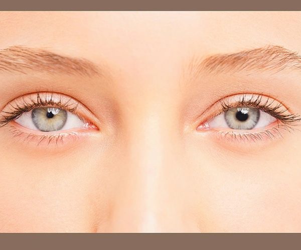
Halfway ptosis (hanging or failure to raise the upper eyelid)
Miosis (choked student)
Facial anhidrosis (nonappearance of perspiring)
The study of disease transmission
Can influence any age, sex, or nationality
Recurrence: roughly 1 for every 6000 people
Neuroanatomy
Horner condition can result from an injury anyplace on the 3-neuron thoughtful pathway providing the eye. The nerve supply begins from the posterolateral nerve center and finishes as the long ciliary nerves that supply the iris dilator and Müller muscles (predominant tarsal muscle).
first request neuron: begins in the nerve center and plummets to the principal neurotransmitter in the cervical spinal line, situated at levels C8–T2 (likewise called the ciliospinal focal point of Budge).
second request neuron: Preganglionic pupillomotor filaments leave the spinal rope at T1, travel through the brachial plexus, over the lung pinnacle, climbing to the predominant cervical ganglion situated close to the point of the mandible and the bifurcation of the normal carotid corridor.
third request neuron: Pupillomotor filaments rise along the inside carotid course and enter the huge sinus where it is in close connection with the abducens nerve (cranial nerve (CN) VI). These strands enter the circle with the ophthalmic branch (V1) of the trigeminal nerve (CN V) through the long ciliary nerves, which innervate the iris dilator and Müller muscles.
Arrangement
first request, or focal, Horner condition: brought about by sores of the thoughtful lots in the mind stem or cervicothoracic spinal string
second request, or preganglionic, Horner condition: brought about by sores that include the spinal string, thoracic outlet, or lung summit. These sores are typically obtained through injury, medical procedure, or harm (e.g., Pancoast growth).
third request, or postganglionic, Horner disorder: brought about by sores of the inner carotid course, like blood vessel analyzation, apoplexy, enormous sinus aneurysm, or wounds procured during carotid conduit stenting
Etiology
Most instances of Horner condition are idiopathic. Of the recognized causes, the etiology relies upon the area of the injury. The causes shift among grown-up and pediatric populaces.
first request condition (focal)
Nerve center:
Stroke
Cancer
Mind stem:
Stroke
Demyelination (e.g., various sclerosis)
Cancer
Spinal string:
Neck injury
Pituitary cancer
Myelitis
Syringomyelia
Demyelination (e.g., different sclerosis)
Arteriovenous deformity (AVM)
Localized necrosis
Arnold-Chiari deformity
Encephalitis
second request disorder (preganglionic)
Apical lung growth (e.g., Pancoast cancer)
Subclavian corridor sores
Mediastinal growths
Cervical rib
Thyroid malignancies
Birth injury with injury to bring down brachial plexus
Mandibular tooth canker
Injuries of the center ear (e.g., intense otitis media)
Iatrogenic (e.g., focal venous catheterization, chest tube position, thoracic medical procedure)
third request disorder (postganglionic)
Inside carotid corridor analyzation
Carotid huge fistula
Injury
Herpes zoster
Nasopharyngeal carcinoma, lymphoma
Bunch or headache migraine
Etiology of Horner disorder in youngsters
Intrinsic (analyzed a month after birth):
Injury to the neck and cerebrum stem during birth
Intrinsic diseases
Neuroblastoma
Idiopathic
Procured:
Neuroblastoma
Rhabdomyosarcoma
Cerebrum stem AVMs
Cerebrum stem growths (glioma)
Demyelination (cerebrum stem)
Carotid conduit apoplexy
Neck injury
Postsurgical (e.g., after jugular cannulation, thoracic medical procedure, or neck a medical procedure)
Pathophysiology
Horner disorder is the aftereffect of the disturbance of the thoughtful inventory to the eye. The manifestations rely upon the area of the sore, and the seriousness relies upon the seriousness of denervation.
Denervation of the nerves that supply the Müller muscle (prevalent tarsal muscle) causes ptosis. Halfway ptosis can happen in light of the fact that the levator palpebrae superioris muscle is unaffected.
Denervation of the nerves that supply the iris dilator muscle causes miosis of the influenced side:
Upon test, anisocoria and a slack of understudy enlargement can be seen.
The light and convenience pupillary reflexes stay ordinary.
Denervation of the nerves that supply the facial perspiration organs causes anhidrosis:
Relies upon the level of interruption of the thoughtful stockpile
Seen in first or second request injuries, however not a noticeable component of third request sores
Clinical Presentation
Exemplary ternion of Horner disorder:
Ptosis
Miosis
Anhidrosis
Visual signs:
Understudies:
Miosis on the influenced side
Anisocoria (distinction in the pupillary size) that is more unmistakable in obscurity.
Enlargement slack (the contracted understudy requires 15–20 seconds longer to widen when a light source is gotten away from the eye.)
Eyelids:
Gentle (< 2 mm) ptosis of the upper eyelid
Reverse ptosis of the lower eyelid (lower cover reprieves at a more significant level than ordinary)
Joined: diminished palpebral opening contrasted with the other eye
Extraocular developments might be influenced in sores of the cerebrum stem or the huge sinus.
Related neurologic signs:
Indications of mind stem injuries:
Ataxia
Diplopia
Lateralized shortcoming
Dizziness
Indications of spinal string (myelopathic) injuries:
Ipsilateral shortcoming
Long lot signs
Entrail and bladder debilitation
Spasticity
Brachial plexopathy: agony and shortcoming in the arm or hand
Cranial neuropathy
In babies and kids, the Harlequin sign (facial flushing) is more evident than anhidrosis.
Iris heterochromia (diverse shaded irides) might be found in kids with inherent Horner disorder. In these cases, the influenced eye has lighter tone.
Conclusion and Management
Clinical analysis
Horner disorder is clinically analyzed by the presence of ptosis and enlargement slack in the influenced eye.
Anhidrosis is likewise generally seen on a similar side as the influenced eye however isn’t required for determination.
Agonizing intense anisocoria is exceptionally reminiscent of Horner condition.
Pharmacologic tests
Effective cocaine can be utilized to affirm the finding of Horner disorder:
Cocaine blocks reuptake of norepinephrine from the synaptic parted and will cause enlargement of the student in the event of a flawless thoughtful pathway. Cocaine has no impact on eyes with impeded thoughtful innervation.
1 hour after the utilization of 2 drops of 10% cocaine, a typical understudy widens more than the Horner student, expanding the level of anisocoria.
Anisocoria ≥ 1 mm after cocaine organization is viewed as a positive outcome.
Effective apraclonidine is utilized as an elective specialist to affirm Horner condition:
A α-adrenergic agonist that causes pupillary enlargement in the Horner understudy and gentle pupillary tightening in the ordinary eye by down-directing norepinephrine
An inversion of anisocoria after the utilization of 2 drops of 0.5% apraclonidine is reminiscent of Horner disorder.
Apraclonidine ought not be utilized in newborn children.
Effective hydroxyamphetamine is utilized to separate first request and second request Horner disorder:
Hydroxyamphetamine causes an arrival of norepinephrine from unblemished adrenergic sensitive spots, causing pupillary widening.
1 hour after the use of 1% hydroxyamphetamine eye drops, widening of the two understudies demonstrates an injury of the first or second request neuron.
On the off chance that the miotic student neglects to enlarge, it shows a third request Horner condition.
Pholedrine can be utilized as an option to hydroxyamphetamine. The test is performed utilizing 1% pholedrine.
Imaging
Imaging is utilized related to clinical trials to affirm the etiology and find the site of the injury.
X-ray of the cerebrum and spinal line is demonstrated in cases with signs characteristic of spinal injuries.
Head CT is likewise informed in cases concerning Horner disorder with a background marked by or clinical signs recommending stroke.
Chest X-beam followed by a CT check should be acted in patients when aspiratory danger is suspected.
The executives
Relies upon the hidden reason
Brief acknowledgment of the condition and determination of the basic etiology is important to decrease deteriorating of the condition.
The intense difficult beginning of Horner disorder ought to be viewed as a health related crisis, as it may demonstrate a carotid supply route analyzation.
Vascular careful attention is important in instances of carotid corridor analyzation.
Neurologic consideration ought to be given in instances of Horner condition identified with aneurysms.
Differential Diagnosis
Adie student: confusion of parasympathetic denervation of the understudy that outcomes in helpless light narrowing yet better convenience. Adie understudy is generally idiopathic. The influenced understudy at first shows up strangely widened very still and has a poor pupillary narrowing in splendid light. Patients gripe of trouble adjusting to dull conditions and photophobia. No treatment is required, however skin physostigmine might give alleviation.
Argyll-Robertson student: portrayed by little and sporadic understudies that choke energetically to approach targets yet respond with almost no tightening to light. Argyll-Robertson student is oftentimes connected with iris decay and is a profoundly explicit indication of neurosyphilis and is treated by dealing with the basic reason.
Constant front uveitis: condition that influences the foremost uvea, iris, and ciliary body. The side effects of uveitis are agony, redness, and photophobia. The indications react well to mitigating drugs. Ongoing foremost uveitis happens in relationship with other constant fiery conditions.
Pupillary sphincter tear: happens due to injury and can cause one-sided or two-sided mydriasis. Patients report glare, haloes in splendidly lit conditions, and inconvenience perusing. Patients with atonic, mydriatic understudies report more awful frown around evening time and failure to zero in on close to objects. The tear requires careful fix.
Aponeurotic ptosis: most normal sort of obtained ptosis. Aponeurotic ptosis happens as a result of outstretching of the levator muscle and is generally found in older patients. Patients generally present with lopsided ptosis or halfway vision misfortune. Careful treatment is required.
Visual myasthenia: visual portrayal of myasthenia gravis. Visual myasthenia brings about visual muscle exhaustion and shortcoming. In myasthenia, antibodies that block the acetylcholine receptor are created, bringing about muscle weariness and loss of motion sometimes. Analysis depends on an edrophonium sodium test. Treatment includes anticholinesterase specialists.


